
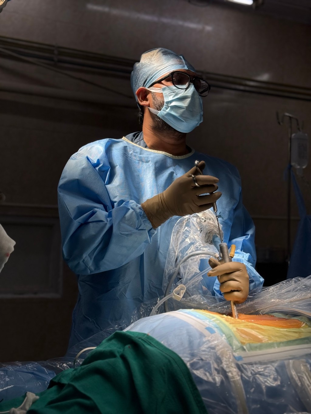
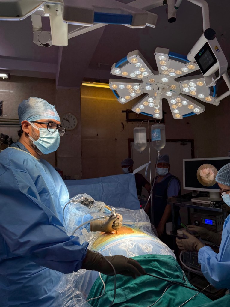
I remember my first endoscopic discectomy like a surgeon remembers their first solo flight.
The working channel: 7mm. The field of view: unfamiliar, disorienting at first — a world seen through a narrow window, lit from within, magnified beyond anything open surgery had prepared me for. The disc fragment: sitting precisely where the MRI said it would be, pressing against a nerve root that had been causing a 52-year-old banker debilitating leg pain for seven months.
I removed it. The nerve moved. It literally moved — decompressed, freed, visibly relaxed.
That moment changed how I think about spine surgery permanently.
This blog is not for patients. It is for the physiotherapist who keeps seeing the same patients cycle back. For the GP who isn’t sure when to refer. For the orthopaedic resident who has heard about endoscopic surgery but never seen it. For the young spine surgeon considering whether to invest the years it takes to learn this technique.
This is what 500 cases taught me that no textbook, no cadaveric workshop, and no fellowship curriculum fully prepared me for.
Lesson 1 —
The Learning Curve Is Real, But It Is Not What You Think
Every paper on endoscopic spine surgery mentions the learning curve. Most cite 20–30 cases before competence, 50–70 before confidence.
What they don’t tell you is that the learning curve is not primarily technical. The optics, the angulation, the water irrigation system — these become second nature faster than expected, especially for surgeons who have trained in arthroscopy or other endoscopic disciplines.
The real learning curve is cognitive.
You are unlearning open surgery intuition and rebuilding spatial understanding from scratch. In open surgery, you orient by what you see directly. In endoscopic surgery, you orient by what you feel through the instruments, what you see on the monitor, and what you have memorised from the pre-operative imaging — simultaneously, in real time, in a working corridor the width of your thumb.
The surgeons who struggle longest are not the ones with poor hands. They are the ones who keep trying to think like open surgeons inside an endoscopic workflow.
The paradigm shift is the curriculum. Not the technique.
Lesson 2 — Patient Selection Is Where the Operation Actually Begins
I have seen endoscopic surgery fail — not in the operating room, but in the outpatient clinic, six weeks before the procedure.
The failure was in patient selection.
Endoscopic spine surgery is not a universal upgrade over open surgery. It is a specific tool with specific indications, and deploying it outside those indications produces predictable disappointment — for the patient and for the surgeon.
My current selection framework, refined over years:
Strong indications for endoscopic approach:
• Single level soft disc herniation (lumbar and cervical)
• Lateral recess stenosis with unilateral radiculopathy
• Foraminal stenosis, particularly in the elderly where minimising soft tissue disruption reduces rehabilitation burden significantly
• Recurrent disc herniation at previously operated levels — where endoscopic access avoids the scarred tissue planes of prior open surgery
Where I pause and think carefully:
• Multi-level disease requiring decompression at contiguous levels
• Significant instability requiring fusion — endoscopy decompresses; it does not stabilise
• Severe central canal stenosis with bilateral symptoms — bilateral endoscopic decompression is possible, but requires advanced skill and honest assessment of your own capability
• Patients with significant psychological overlay or chronic pain syndrome — no surgical approach resolves what is fundamentally a central sensitisation problem
The surgeon who offers endoscopic surgery to every spine patient is not progressive. They are operating outside the evidence — and eventually, outside the patient’s best interest.
Lesson 3 — The Complication You Fear Most Is Not the One That Humbles You
Ask any endoscopic spine surgeon what they fear, and they will say: dural tear, nerve injury, inadequate decompression requiring revision.
These are real risks. They deserve respect.
But the complication that has taught me the most — the one that humbled me earliest and sharpest — was none of these.
It was inadequate decompression in a patient who left the operating room pain-free.
Let me explain.
In endoscopic surgery, the immediate tactile and visual feedback of decompression can be misleading. The nerve root looks free. The disc fragment is removed. The patient wakes up with dramatically reduced pain — and you feel the quiet satisfaction of a procedure done well.
Then, at six weeks, the radiculopathy returns. Not because of reherniation. Because you decompressed the primary fragment but left a secondary migrated fragment two millimetres cranial that the intraoperative imaging did not catch.
This taught me two things permanently:
First — intraoperative fluoroscopy and endoscopic visualisation must be used together, not interchangeably. One shows you where you are. The other shows you what you are doing. They answer different questions.
Second — a patient leaving the OT pain-free is a data point, not a conclusion. The follow-up at six weeks is where the operation is truly judged.
Lesson 4 — What the Referring Doctor Needs to Know (And Rarely Hears)
I receive referrals from GPs, physiotherapists, neurologists, and general orthopaedic surgeons across Mumbai. I am grateful for every one of them. And I want to be useful to those referrers — which means being honest about what helps me help your patients.
Refer earlier for radiculopathy with neurological deficit.
The window for nerve recovery narrows with time. A patient with foot drop of six weeks duration has a meaningfully better prognosis than one with six months. If motor deficit is present, the referral should not wait for completion of a full conservative trial
The physiotherapy you prescribe matters.
McKenzie-based directional preference therapy for disc pathology, and specific core stabilisation for mechanical back pain, are not interchangeable with generic back exercises.
When physiotherapy fails, I want to know which physiotherapy failed — because the answer changes my surgical planning and my post-operative rehabilitation prescription.
Imaging without clinical correlation creates anxiety, not clarity.
Please resist the pressure — from patients and from radiologists’ incidental findings — to refer asymptomatic or minimally symptomatic patients for surgical consultation based on imaging alone. It creates false urgency, undermines trust in the medical system, and places the spine surgeon in the uncomfortable position of explaining why a “terrible” MRI may require no intervention.
Send me the patient. Send me the story. The MRI is supporting evidence.
Lesson 5 — India Is Not Behind. India Is Different.
This is perhaps the most important perspective I can offer.
There is a tendency among Indian spine surgeons — particularly those trained abroad — to benchmark Indian spine surgery practice against Western or East Asian standards and find ourselves lacking. Longer delays to presentation. Less structured rehabilitation infrastructure. More socioeconomic constraints on treatment decisions.
I want to reframe this.
Indian spine surgeons operating in busy urban centres like Mumbai are, in many ways, accumulating case experience and surgical volume that would take twice as long in a Western practice. The diversity of pathology — the range of ages, body types, occupational demands, and chronicity of disease — is extraordinary.
What we lack is not skill or volume. What we lack is structured outcomes data collection, peer-reviewed publication of our results, and the clinical networks that turn individual surgical experience into collective surgical knowledge.
The endoscopic spine surgery community in India is growing rapidly. The technical capability exists. What we must now build — together, as a community — is the evidence base that reflects what we are actually achieving in our operating rooms.
Because what I see in my OT in Chembur/ Parel deserves to be in the literature.
And what you are seeing in yours does too.
A Final Thought for the Young Spine Surgeon Reading This
Learn endoscopic surgery. Not because it is fashionable. Not because patients ask for it by name. Learn it because it will make you a better spine surgeon regardless of which approach you ultimately use.
The discipline of working through a small corridor forces a precision of anatomy, a respect for tissue planes, and a humility about what is truly necessary that open surgery — with its generous exposure and forgiving access — does not always demand.
The best open spine surgeons I know think endoscopically even when their hands are working openly. They take only what they need. They disturb only what must be disturbed. They leave the body as close to how they found it as the pathology allows.
That, ultimately, is what this technique taught me.
Not a new way to operate. A new standard for what operating well actually means.
📍 Dr. Om Patil — Endoscopic Spine Surgeon, Mumbai
For referrals, second opinions, or surgical consultations:
📞 +91 98191 95885
505, Signature Business Park
Postal Colony, Chembur East
Mumbai — 400071
🌐 http://www.spinewala.com
🌐 http://www.spinesurgerymumbai.com
Www.orthdocsurgeon.com
Dr. Om Patil is an endoscopic spine surgeon in Mumbai with a focused practice in minimally invasive and endoscopic techniques for lumbar and cervical spine pathology. He writes on surgical philosophy, patient education, and the evolving landscape of spine care in India.
 Spine Surgeons Notes
Leave a comment