

When we discuss the health impacts of menopause, vasomotor symptoms like hot flashes dominate the conversation. But the most profound architectural transformation is happening silently within the skeleton—specifically, the spine.

Post-menopausal women experience a rapid acceleration of bone remodeling that disproportionately impacts vertebral structures. To preserve long-term mobility and independence, we must bridge the gap between endocrine science and practical musculoskeletal biomechanics.
The Triad of Decline: Why the Spine is Vulnerable
The post-menopausal spine faces a trifecta of structural challenges driven by the sharp decline in circulating 17\beta\text{-estradiol} (estrogen). Estrogen acts as a primary regulator of bone and tissue homeostasis; its withdrawal triggers three distinct degenerative pathways:
- Accelerated Trabecular Bone Loss: Estrogen suppresses osteoclast (bone-resorbing cell) activity by regulating the RANKL/OPG signaling pathway. Without it, bone breakdown goes unchecked. Because vertebral bodies are highly composed of metabolic-rich trabecular (spongy) bone, the spine loses density faster than long bones—sometimes declining by 3% to 5% annually in the immediate years post-menopause.
- Intervertebral Disc Degeneration (IDD): Estrogen receptors are present within the nucleus pulposus and annulus fibrosus of spinal discs. The loss of estrogen decreases proteoglycan synthesis, leading to disc dehydration, loss of disc height, and reduced mechanical shock absorption.
- Paraspinal Sarcopenia: The age-related loss of skeletal muscle mass accelerates sharply after menopause. As the deep stabilizing muscles of the spine—such as the multifidus and erector spinae—weaken, the structural load shifts from muscular support directly onto an already compromised skeletal framework.
Prevention: The Biomechanical and Nutritional Defense
Preventing structural failure (such as vertebral compression fractures) requires moving past general lifestyle advice and focusing on targeted mechanical loading and bone-matrix biochemistry.
1. Targeted Mechanical Loading (Progressive Overload)
Generic, low-impact cardiovascular exercise like walking or swimming is excellent for metabolic health, but it is insufficient to stimulate bone deposition (osteogenesis). The skeleton responds strictly to high-rate mechanical strain.
- Axial Loading: Under professional supervision, integrating compound movements that load the spine vertically (such as modified deadlifts or goblet squats) triggers the mechanoreceptors in osteocytes to signal new bone formation.
- Back Extension Prioritization: Strengthening the erector spinae and deep core stabilization complexes (e.g., tailored Pilates frameworks) reduces the anterior shearing forces on the vertebral bodies, protecting against hyperkyphosis (“dowager’s hump”).
2. The Micronutrient Framework
Bone matrix formation relies on specific biochemical co-factors. Supplementation should bridge nutritional gaps to support this process:
- Calcium: 1,200 mg/day (ideally prioritized through dietary sources like fortified foods, leafy greens, and dairy; supplemented if necessary).
- Vitamin D3: 800–2,000 IU/day (titrated to maintain optimal serum 25(OH)D levels) to enable active intestinal calcium absorption.
- Vitamin K2 (MK-7): 90–120 mcg/day to activate osteocalcin, the protein responsible for binding calcium directly to the hydroxyapatite bone matrix rather than soft tissues.
- Dietary Protein: 1.0–1.2 g/kg of body weight to support paraspinal muscle mass and the collagenous framework of the bone matrix.
Clinical Treatment Pathways
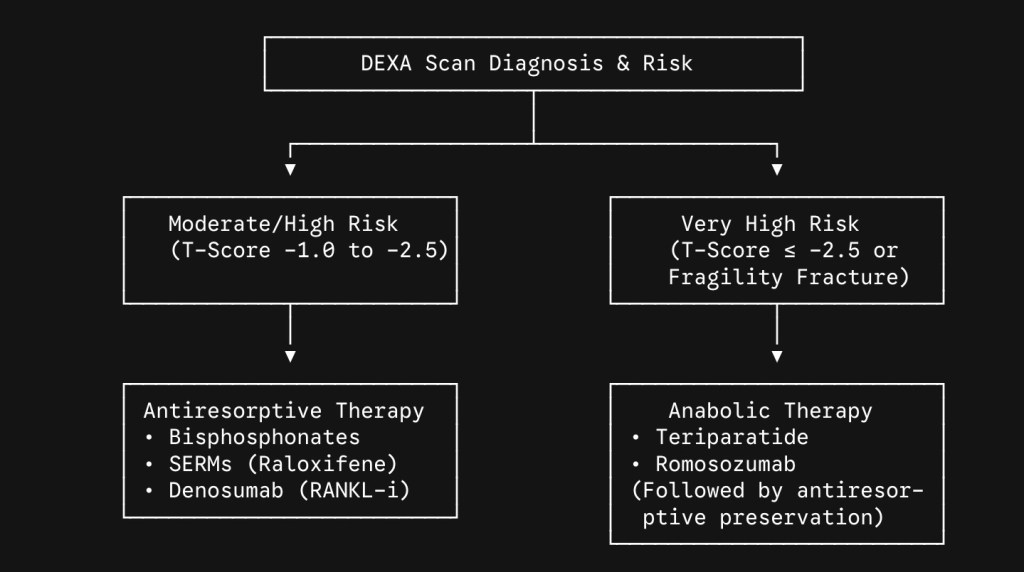
When a Dual-Energy X-ray Absorptiometry (DEXA) scan reveals osteoporosis (T-score less than or equal to -2.5) or a fragility fracture occurs, mechanical and nutritional interventions must be paired with pharmacological therapy.
Antiresorptive Agents (Bone-Preserving)
- Bisphosphonates (e.g., Alendronate, Zoledronic Acid): These compounds bind to hydroxyapatite crystals and inhibit osteoclast-mediated bone resorption, significantly reducing vertebral fracture risk.
- Denosumab: A monoclonal antibody that targets RANKL, preventing osteoclast maturation. Clinical note: Discontinuation requires an immediate transition to an alternative therapy to avoid a rebound increase in bone resorption.
- SERMs (e.g., Raloxifene): Act as selective estrogen agonists in bone tissue to reduce resorption while acting as antagonists in breast tissue.
Anabolic Agents (Bone-Building)
- PTH/PTHrP Analogs (e.g., Teriparatide, Abaloparatide): Intermittent administration stimulates osteoblast (bone-building cell) activity over osteoclasts, actively building new cortical and trabecular architecture.
- Sclerostin Inhibitors (e.g., Romosozumab): A dual-action monoclonal antibody that simultaneously accelerates bone formation while decreasing bone resorption.
💡 The Bottom Line for Clinicians & Leaders: Do not wait for a fragility fracture to occur before assessing a patient’s skeletal health. Proactive baseline DEXA screening, early implementation of heavy resistance training, and timely pharmacological intervention are essential to maintaining long-term physical autonomy and spinal health post-menopause.
#WomensHealth #Menopause #SpineHealth #Orthopedics #PhysicalTherapy #Osteoporosis Prevention #HealthyAging
 Spine Surgeons Notes
Leave a comment